Page 26 Guide to Pain Management in Low-Resource Settings
P. 26
14 Nilesh B. Patel
and (2) larger diameter, lightly myelinated nerves that Table 1
conduct nerve impulses faster (20 m/sec = 72 km/h) Selected chemical substances released with stimuli
termed Aδ fi bers. Th e C-fi ber nociceptors respond poly- suffi cient to cause tissue damage
modally to thermal, mechanical, and chemical stimuli; Substance Source
and the Aδ-fi ber nociceptors are of two types and re- Potassium Damaged cells
spond to mechanical and mechanothermal stimuli. It Serotonin Platelets
is well known that the sensation of pain is made up of Bradykinin Plasma
two categories—an initial fast, sharp (“epicritic”) pain Histamine Mast cells
and a later slow, dull, long lasting (“protopathic”) pain. Prostaglandins Damaged cells
Th is pattern is explained by the diff erence in the speed Leukotrienes Damaged cells
of propagation of nerve impulses in the two nerve fi ber Substance P Primary nerve aff erents
types described above. Th e neuronal impulses in fast-
Hypersensitivity may be diagnosed by taking
conducting Aδ-fi ber nociceptors produce the sensation
history and by careful examination. Certain conditions
of the sharp, fast pain, while the slower C-fi ber nocicep-
may be discriminated:
tors produce the sensation of the delayed, dull pain.
a) Allodynia: Pain due to a stimulus that does not
Peripheral activation of the nociceptors (trans-
normally provoke pain, e.g., pain caused by a T-shirt in
duction) is modulated by a number of chemical sub-
patients with postherpetic neuralgia.
stances, which are produced or released when there is
b) Dysesthesia: An unpleasant abnormal sensation,
cellular damage (Table 1). Th ese mediators infl uence the
whether spontaneous or evoked. (Note: a dysesthesia
degree of nerve activity and, hence, the intensity of the
should always be unpleasant, while paresthesia should
pain sensation. Repeated stimulation typically causes
not be unpleasant; e.g., in patients with diabetic poly-
sensitization of peripheral nerve fi bers, causing lower-
neuropathy or vitamin B defi ciency.)
ing of pain thresholds and spontaneous pain, a mecha- 1
c) Hyperalgesia: An increased response to a stimu-
nism that can be experienced as cutaneous hypersensi-
lus that is normally painful. (Note: hyperalgesia refl ects
tivity, e.g., in skin areas with sunburn.
increased pain on suprathreshold stimulation; e.g., in
patients with neuropathies as a consequence of pertur-
bation of the nociceptive system with peripheral and/or
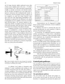
Released by
tissue damage:
Skin Bradykinin central sensitization.)
K+ d) Hyperesthesia: Increased sensitivity to stimula-
Prostaglandins
tion, excluding the special senses, e.g., increased cuta-
C fibers
neous sensibility to thermal sensation without pain.
Aδ fibers
Histamine
Injury To spinal cord With the knowledge of pain pathways and sen-
sitization mechanisms, therapeutic strategies to inter-
act specifi cally with the pain generation mechanisms
can be developed.
Mast
Cell
Fig. 1. Some chemicals released by tissue damage that stimulates Central pain pathways
nociceptors. In addition release of substance-P, along with hista-
mine, produce vasodilation and swelling.
Th e spinothalamic pathway and the trigeminal pathway
are the major nerve routes for the transmission of pain
In addition, local release of chemicals such
and normal temperature information from the body and
substance P causes vasodilation and swelling as well
face to the brain. Visceral organs have only C-fi ber noci-
as release of histamine from the mast cells, further in-
ceptive nerves, and thus there is no refl ex action due to
creasing vasodilation. Th is complex chemical signaling
visceral organ pain.
protects the injured area by producing behaviors that
keep that area away from mechanical or other stimuli. Th e spinothalamic pathway
Promotion of healing and protection against infection The nerve fibers from the dorsal root ganglia en-
are aided by the increased blood fl ow and infl ammation ter the spinal cord through the dorsal root and send
(the “protective function of pain”). branches 1–2 segments up and down the spinal cord