Page 51 Acute Pain Management
P. 51
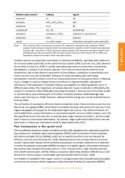
Metabotropic receptor Subtype Ligand
histamine H 1 HA
serotonin 5HT 1A , 5HT 4 , 5HT 2A 5HT
bradykinin B I , B 2 BK
cannabinoid CB 1 , CB 2 anandamide
tachykinin neurokinin‐1 (NK 1 ) substance P, neurokinin A
proteinase PAR 1‐4 protease
opioid mu, delta, kappa enkephalin, dynorphin, beta‐endorphin
Notes: 5HT: serotonin; ASIC: acid sensing ion channel; ATP: adenosine triphosphate; BK: bradykinin; DRASIC:
subtype of acid sensing ion channel; iGluR: ionotropic glutamate receptor; mGluP: metabotropic glutamate
receptor; NK1: neurokinin‐1; P2X3: purinergic receptor subtype; PAR: proteinase‐activated receptor; PGE 2:
prostaglandin E2; PGI 2: prostacyclin; TRP: transient receptor potential. Others (eg H1, EP1‐4, TRPV 2) are
designated subtypes of receptors rather than abbreviations.
Sodium channels are important modulators of neuronal excitability, signalling and conduction CHAPTER 1
of neuronal action potentials to the central nervous system (CNS) (Cummins et al, 2007; Momin &
Wood, 2008; Dib‐Hajj et al, 2009). A rapidly inactivating fast sodium current that is blocked by
tetrodotoxin is present in all sensory neurons. This is the principal site of action for local
anaesthetics, but as the channel is present in all nerve fibres, conduction in sympathetic and
motor neurons may also be blocked. Subtypes of slowly activating and inactivating
tetrodotoxin‐resistant sodium currents are selectively present on nociceptive fibres. Following
injury, changes in sodium channel kinetics contribute to hyperexcitability, and specific
alterations in the expression of sodium channels (upregulation or downregulation) occur in
different pain states. The importance of sodium channels in pain sensitivity is reflected by the
impact of mutations in the SCN9A gene encoding the Na(v)1.7 channel: loss‐of‐function results
in insensitivity to pain whereas gain‐of‐function mutations produce erythromelalgia and
severe pain (Dib‐Hajj et al, 2008). However, subtype‐selective drugs are not yet available (Momin
& Wood, 2008).
The cell bodies of nociceptive afferents that innervate the trunk, limbs and viscera are found in
the dorsal root ganglia (DRG), while those innervating the head, oral cavity and neck are in the
trigeminal ganglia and project to the brainstem trigeminal nucleus. The central terminals of C
and A‐delta fibres convey information to nociceptive‐specific neurons within laminae I and II of
the superficial dorsal horn and also to wide dynamic range neurons in lamina V, which encode
both innocuous and noxious information. By contrast, large myelinated A‐beta fibres transmit
light touch or innocuous mechanical stimuli to deep laminae III and IV.
Pain transmission in the spinal cord
Primary afferent terminals contain excitatory amino acids (eg glutamate, aspartate), peptides
(eg substance P, calcitonin gene‐related peptide [CGRP]) and neurotrophic factors (eg brain‐
derived neurotrophic factor [BDNF]), which act as neurotransmitters and are released by
different intensity stimuli (Sandkuhler, 2009). Depolarisation of the primary afferent terminal
results in glutamate release, which activates postsynaptic ionotropic alpha‐amino‐3‐hydroxyl‐
5‐methyl‐4‐isoxazole‐propionate (AMPA) receptors and rapidly signals information relating to
the location and intensity of noxious stimuli. In this ‘normal mode’ a high intensity stimulus
elicits brief localised pain, and the stimulus‐response relationship between afferent input and
dorsal horn neuron output is predictable and reproducible (Woolf & Salter, 2000).
Summation of repeated C‐fibre inputs results in a progressively more depolarised postsynaptic
membrane and removal of the magnesium block from the N‐methyl‐D‐aspartate (NMDA)
Acute pain management: scientific evidence 3