Page 53 Acute Pain Management
P. 53
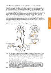
(ie the site and type of painful stimulus). The spinoreticular (spinoparabrachial) and
spinomesencephalic tracts project to the medulla and brainstem and are important for
integrating nociceptive information with arousal, homeostatic and autonomic responses as
well as projecting to central areas mediating the emotional or affective component of pain.
The spinoparabrachial pathway originates from superficial dorsal horn lamina I neurons that
express the NK 1 receptor and projects to the ventromedial hypothalamus and central nucleus
of the amygdala. Multiple further connections include those with cortical areas involved in the
affective and motivational components of pain (eg anterior cingulate cortex, insular and
prefrontal cortex), projections back to the periaqueductal grey (PAG) region of the midbrain
and rostroventromedial medulla (RVM), which are crucial for fight or flight responses and
stress‐induced analgesia, and projections to the reticular formation, which are important for
the regulation of descending pathways to the spinal cord (see Figure 1.1) (Hunt & Mantyh, 2001;
Tracey & Mantyh, 2007; Tracey, 2008).
Figure 1.1 The main ascending and descending spinal pain pathways CHAPTER 1
ASCENDING DESCENDING
PATHWAYS PATHWAYS
cc cc
Hip Hip
Po Po
ic ic
VPM/VPL VPM/VPL
Ce VMH Ce VMH
PAG BRAINSTEM
opioids
clonidine
A7 N 2O
A TCA
5
PB
bc LC
V RVM
PERIPHERY
NSAIDs NERVE CONDUCTION
Py
opioids local anaesthetics
SPINAL CORD
opioids
clonidine
DRG
ketamine
TCA
NSAIDs
Notes: (a) There are 2 primary ascending nociceptive pathways. The spinoparabrachial pathway (red) originates
from the superficial dorsal horn and feeds areas of the brain concerned with affect. The spinothalamic
pathway (blue) originates from deeper in the dorsal horn (lamina V) after receiving input from the
superficial dorsal horn and predominantly distributes nociceptive information to areas of the cortex
concerned with discrimination.
(b) The descending pathway highlighted originates from the amygdala and hypothalamus and terminates in
the PAG. Neurons project from here to the lower brainstem and control many of the antinociceptive and
autonomic responses that follow noxious stimulation.
Other less prominent pathways are not illustrated.
The site of action of some commonly utilised analgesics are included.
Legend A: adrenergic nucleus; bc: brachium conjunctivum; cc: corpus collosum; Ce: central nucleus of the
amygdala; DRG: dorsal root ganglion; Hip: hippocampus; ic: internal capsule; LC: locus coeruleus; PAG:
periaqueductal grey; PB: parabrachial area; Po: posterior group of thalamic nuclei; Py: pyramidal tract;
RVM: rostroventrmedial medulla; V: ventricle; VMH: ventral medial nucleus of the hypothalamus; VPL:
ventral posterolateral nucleus of the thalamus; VPM: ventral posteromedial nucleus of the thalamus
Source: Modified from Hunt (Hunt & Mantyh, 2001).
Acute pain management: scientific evidence 5